"I Survived. I Lived. Then I Woke Up."

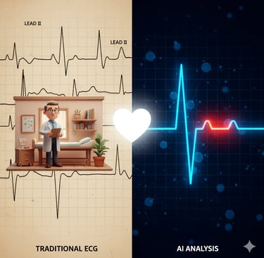
Rural Hearts, Smart Tech: How WVU’s AI Turns Simple ECGs into Early Warnings
West Virginia University scientists trained AI on 55,000+ Appalachian patient ECGs to spot signs of heart failure—using low-cost tools that rural clinics already have. Here’s why that matters, how it works, and what comes next.
Christopher J
9/18/20253 min read

The story in a heartbeat
A team at West Virginia University trained artificial intelligence to read ordinary 12-lead ECGs—the sticky-pad test you can do in minutes—and estimate the heart’s ejection fraction, a key measure used to flag heart failure. Instead of depending on pricey echocardiograms and urban hospital datasets, they built and tested models on more than 55,000 West Virginians across 28 hospitals. Translation: smarter screening using tools rural clinics already own. WVU Today+1
What exactly did they build?
The researchers compared a bunch of machine-learning and deep-learning models on ECG signals linked to each patient’s echocardiogram-derived ejection fraction. Deep models won, with a 1-D ResNet architecture posting the strongest results—area under the ROC curve around 0.86 at one clinically meaningful threshold. They also tested which ECG “leads” (the different electrode pairs) mattered most and found specific combinations improved accuracy over any single lead alone. Nature
Why is this a big deal for rural care?
Heart failure hits rural Appalachia especially hard, yet AI tools are often trained on patients from big coastal systems—think Stanford and other urban datasets—then shipped everywhere else. WVU’s team flipped that script by training on Appalachian patients to reduce bias and better reflect local realities like limited specialty access. As project lead Prashnna Gyawali put it, “This is why this work matters”—so people like a typical “Jane Doe” in rural West Virginia get models tuned to their context, not someone else’s. WVU Today
ECG vs. echo: the access advantage
Echocardiography (the usual way to measure ejection fraction) is excellent but costs time, money, and specialized staffing. An ECG is cheap, quick, and everywhere—even in small clinics and urgent care settings. Doctoral researcher Alina Devkota explained that the team trained models to predict ejection fraction straight from ECGs, meeting patients where they already are. If screening works well, clinics could flag risk earlier and fast-track the right people for imaging and treatment. WVU Today
Nerdy bits without the nosebleed
• Data: 55,500 unique patients; ECGs sampled at 500 Hz; patient splits to avoid leakage between train/validation/test.
• Models: traditional (SVM, Random Forest, etc.) versus deep (ResNet, Transformers). ResNet took the crown; Transformers were the runner-up.
• Thresholds: they tested several ejection-fraction cutoffs (35%, 40%, 45%, 50%) to see where models are most reliable; performance was best at the lower (sicker) threshold.
• Leads: no single lead was enough; certain pairings (like I, aVL, aVF, V1 variants) helped, but full 12-lead input performed best. Nature
So…is this ready for your next clinic visit?
Not yet. The WVU team is careful: AI isn’t in clinical practice here today, and reliability, validation across sites, and regulatory approvals matter. But the direction is promising: low-cost, low-friction triage that could “level up” primary care—especially where long wait times and travel make advanced imaging tough. The authors argue that training and validating on the population you intend to serve is non-negotiable for fairness and clinical usefulness. WVU Today+1
Context from the wider field
Other groups have shown you can detect reduced ejection fraction from noninvasive signals (including ECG and even heart sounds), but models often rely on urban datasets. WVU’s contribution is scale and local relevance: a state-wide, de-identified dataset purpose-built for Appalachia, plus systematic comparisons across model types and lead combinations. That’s how you move from “cool demo” to “clinically sensible.” PMC+1
What experts are saying (short and sweet)
• “Ensure people like Jane receive accurate diagnoses”—Gyawali’s emphasis on population-specific training. WVU Today
• Devkota’s rationale: ECGs are inexpensive and everywhere, so pairing them with AI could help the very communities with the highest burden of disease. WVU Today
Caveats worth keeping in your pocket
This is still retrospective analysis; prospective, multi-center trials are the next checkpoint. Model performance can wobble if the patient mix, device settings, or prevalence shifts. And even a strong AUROC doesn’t tell you how a tool impacts outcomes—like fewer hospitalizations or faster therapy. Those require careful implementation studies (and yes, lots of IRB forms). Nature
What this means for you (and your heart)
If you live far from a cardiology hub, this research hints at a future where a routine ECG plus AI could flag problems early—before symptoms escalate. In the meantime, the basics still win most days: move your body, watch blood pressure and sodium, keep follow-ups, and don’t ignore new shortness of breath or swelling. As I share in my recovery story, progress often comes from pairing humble daily tools with relentless consistency. AI is just another tool—powerful, yes, but most powerful when it meets patients where they are and nudges action sooner rather than later.

Do you have a life changing story and want to help others with your experience and inspiration. Please DM me or Send me an email at the links below
Contact Me
© 2025. All rights reserved.
Privacy Policy